Home » Without Label » Abdominal Anatomy - Abdominal Anatomy Medical Illustration - avenue-v / The abdominal aorta enters the abdomen through the diaphragm at the level of the twelfth thoracic vertebre and continues to just below the umbilical area, where it splits into the right and left common iliac arteries.
Abdominal Anatomy - Abdominal Anatomy Medical Illustration - avenue-v / The abdominal aorta enters the abdomen through the diaphragm at the level of the twelfth thoracic vertebre and continues to just below the umbilical area, where it splits into the right and left common iliac arteries.
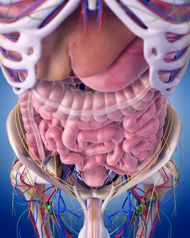
Abdominal Anatomy - Abdominal Anatomy Medical Illustration - avenue-v / The abdominal aorta enters the abdomen through the diaphragm at the level of the twelfth thoracic vertebre and continues to just below the umbilical area, where it splits into the right and left common iliac arteries.. The abdomen is the part of the body that contains all of the structures between the thorax (chest) and the pelvis, and is separated from the thorax via the diaphragm. The major organs of the abdomen include the small intestine, large intestine, and stomach. The abdomen contains all the digestive organs, including the stomach, small and large intestines, pancreas, liver, and gallbladder. The abdominal cavity is the part of the body that houses the stomach, liver, pancreas, kidneys, gallbladder, spleen, and the large and small intestines.the diaphragm marks the top of the abdomen and the horizontal line at the level of the top of the pelvis marks the bottom. The component of the urinary system, kidney and the ureter.
The regions occupied by stomach are epigastric, umbilical and hypochondriac regions. It also contains the spleen. This anatomy section promotes the use of the terminologia anatomica , the international standard of anatomical nomenclature. This mri abdomen axial cross sectional anatomy tool is absolutely free to use. However, the imaging test may be used to diagnose or rule out many other health conditions.
The abdominal anatomy stock illustration. Illustration of ... from thumbs.dreamstime.com Stomach is a muscular bag forming the most distensible part of the human digestive system. Together, these three turn nutrients into usable energy, as well as help dispose of solid waste. It also contains the spleen. Abdominal anatomy includes a major element of the gastrointestinal, system, the caudal end of the oesophagus, stomach, large and small intestine, liver, pancreas and the gallbladder. It is an artery, meaning that it carries blood away from the heart. Abdominal and pelvic anatomy encompasses the anatomy of all structures of the abdominal and pelvic cavities. The aorta is the largest blood vessel in the body. Terms in this set (94) what is the abdomen.
It's the preferred screening method for an abdominal aortic aneurysm, a weakened, bulging spot in the abdominal aorta — the major blood vessel that supplies blood to the body.
It is the long, flat muscle that extends vertically between the pubis and the fifth, sixth, and seventh ribs. Together, these three turn nutrients into usable energy, as well as help dispose of solid waste. It is bounded superiorly by the xiphoid process and costal margins, posteriorly by the vertebral column and inferiorly by the pelvic bones and inguinal ligament. Boundaries of the abdomen (4) anterior abdominal wall (anterolateral) diaphragm (superior) pelvic inlet (inferior) This area contains various subdivisions. An abdominal ultrasound is done to view structures inside the abdomen. However, the imaging test may be used to diagnose or rule out many other health conditions. The major organs of the abdomen include the small intestine, large intestine, and stomach. In anatomy and physiology, you'll learn how to divide the abdomen into nine different regions and four different quadrants. The aorta is the largest blood vessel in the body. We're going to take apart a plastic anatomy model and see what we can find in the abdomen. The abdominal (peritoneal) cavity is an area that normally only contains a small amount of peritoneal fluid, however can become a potential space for pathology. Inferiorly the abdomen is open to the pelvis, communicating through the superior pelvic aperture (pelvic inlet).
Abdomen, in human anatomy, the body cavity lying between the chest or thorax above and the pelvis below and from the spine in the back to the wall of abdominal muscles in the front. This mri abdomen axial cross sectional anatomy tool is absolutely free to use. The abdomen (colloquially called the belly, tummy, midriff or stomach) is the part of the body between the thorax (chest) and pelvis, in humans and in other vertebrates.the abdomen is the front part of the abdominal segment of the trunk.the area occupied by the abdomen is called the abdominal cavity.in arthropods it is the posterior tagma of the body; It follows the thorax or cephalothorax. It is the long, flat muscle that extends vertically between the pubis and the fifth, sixth, and seventh ribs.
Vintage 1950's Frohse Chest & Abdomen Viscera Human ... from cdn.shopify.com The abdomen contains many vital organs: It also contains the spleen. Boundaries of the abdomen (4) anterior abdominal wall (anterolateral) diaphragm (superior) pelvic inlet (inferior) Abdominal anatomy includes a major element of the gastrointestinal, system, the caudal end of the oesophagus, stomach, large and small intestine, liver, pancreas and the gallbladder. The abdominal wall surrounds the abdominal cavity, providing it with flexible coverage and protecting the internal organs from damage. Abdomen anatomy the abdomen is comprised primarily of the digestive tract and other accessory organs which assist in digestion, the urinary system, spleen, and the abdominal muscles (shown below). This area contains various subdivisions. We'll identify as many organs as we can, see how they fit into the.
The major organs of the abdomen include the small intestine, large intestine, and stomach.
The abdomen (colloquially called the belly, tummy, midriff or stomach) is the part of the body between the thorax (chest) and pelvis, in humans and in other vertebrates.the abdomen is the front part of the abdominal segment of the trunk.the area occupied by the abdomen is called the abdominal cavity.in arthropods it is the posterior tagma of the body; The main areas of the abdomen include the abdominal cavity, calot's triangle, the peritoneum, the inguinal canal, and hesselbach's triangle. Together, these three turn nutrients into usable energy, as well as help dispose of solid waste. It also contains the spleen. Boundaries of the abdomen (4) anterior abdominal wall (anterolateral) diaphragm (superior) pelvic inlet (inferior) Together, these three turn nutrients into usable energy, as well as help dispose of solid waste. Assoc prof craig hacking and dr tim luijkx et al. Abdomen, in human anatomy, the body cavity lying between the chest or thorax above and the pelvis below and from the spine in the back to the wall of abdominal muscles in the front. The abdominal (peritoneal) cavity is an area that normally only contains a small amount of peritoneal fluid, however can become a potential space for pathology. The abdominal wall surrounds the abdominal cavity, providing it with flexible coverage and protecting the internal organs from damage. Abdominal computed tomography (ct) is a type of medical imaging procedure used to diagnose and monitor internal stomach issues, like cancer, bowel obstruction, and abdominal pain. The major organs of the abdomen include the small intestine, large intestine, and stomach. We'll identify as many organs as we can, see how they fit into the.
This anatomy section promotes the use of the terminologia anatomica , the international standard of anatomical nomenclature. This area contains various subdivisions. In anatomy and physiology, you'll learn how to divide the abdomen into nine different regions and four different quadrants. An abdominal ultrasound is done to view structures inside the abdomen. It also contains the spleen.
Bodybuilding Anatomy - Meet Your Muscles from bodybuilding-wizard.com The abdomen contains all the digestive organs, including the stomach, small and large intestines, pancreas, liver, and gallbladder. It is bounded superiorly by the xiphoid process and costal margins, posteriorly by the vertebral column and inferiorly by the pelvic bones and inguinal ligament. However, the imaging test may be used to diagnose or rule out many other health conditions. The rectus abdominis connects to the xiphoid process, a bony landmark at the bottom of the sternum. The abdominal cavity is the part of the body that houses the stomach, liver, pancreas, kidneys, gallbladder, spleen, and the large and small intestines.the diaphragm marks the top of the abdomen and the horizontal line at the level of the top of the pelvis marks the bottom. These organs are held together loosely by connecting tissues. The abdominal (peritoneal) cavity is an area that normally only contains a small amount of peritoneal fluid, however can become a potential space for pathology. The abdomen is the part of the body that contains all of the structures between the thorax (chest) and the pelvis, and is separated from the thorax via the diaphragm.
The aorta is the largest blood vessel in the body.
The component of the urinary system, kidney and the ureter. The abdominal wall surrounds the abdominal cavity, providing it with flexible coverage and protecting the internal organs from damage. The abdomen contains many vital organs: In anatomy and physiology, you'll learn how to divide the abdomen into nine different regions and four different quadrants. An abdominal ultrasound is done to view structures inside the abdomen. It is the long, flat muscle that extends vertically between the pubis and the fifth, sixth, and seventh ribs. The major organs of the abdomen include the small intestine, large intestine, and stomach. However, the imaging test may be used to diagnose or rule out many other health conditions. The abdomen is the body region found between the thorax and the pelvis. The abdominal aorta enters the abdomen through the diaphragm at the level of the twelfth thoracic vertebre and continues to just below the umbilical area, where it splits into the right and left common iliac arteries. This mri abdomen axial cross sectional anatomy tool is absolutely free to use. Part of the trunk between thorax and pelvis. Abdomen, in human anatomy, the body cavity lying between the chest or thorax above and the pelvis below and from the spine in the back to the wall of abdominal muscles in the front.